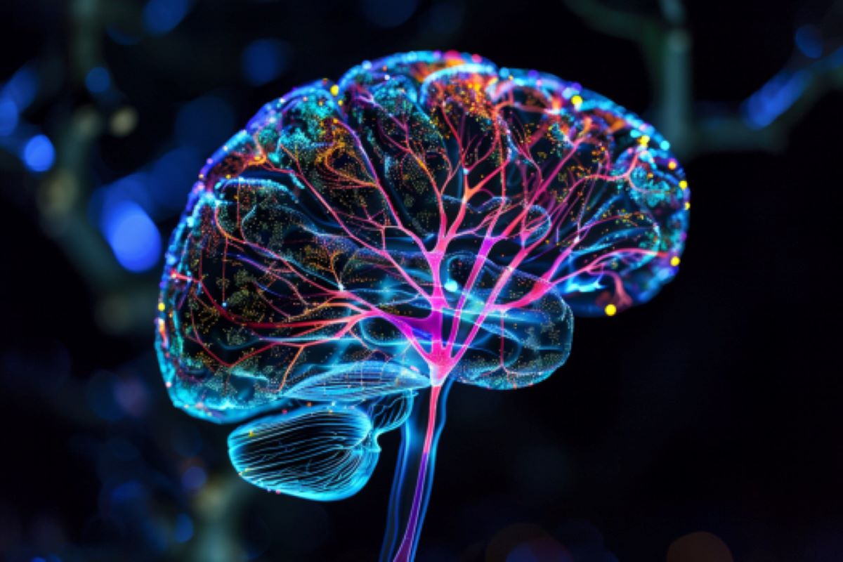
Summary: Researchers have introduced a new bioluminescence detection technique to image deep brain structures, bypassing the challenge of light scattering. This method involves engineering the brain’s blood vessels to dilate in response to light, which can then be monitored using MRI.
By sensitizing the brain’s vasculature to act as light detectors, the researchers effectively transformed these blood vessels into a “three-dimensional chamber.” This breakthrough opens up new possibilities for studying brain function and mapping changes in gene expression in unprecedented detail.
Key facts:
- Bioluminescence as a tool: Commonly used to track cellular processes, bioluminescence involves tagging cells with proteins such as luciferase that emit light, allowing researchers to visualize biological phenomena.
- An innovative detection method: The developed technique turns blood vessels into bioluminescence detectors, using MRI to image the dilations caused by light-responsive proteins, overcoming the problem of light scattering in deep tissues.
- Potential applications: This method could greatly improve research opportunities in neuroscience, enabling detailed mapping of gene expression and cellular interactions in the brain, potentially leading to advances in the understanding of neurological diseases and brain function.
source: MIT
Scientists often tag cells with proteins that glow, allowing them to track tumor growth or measure changes in gene expression that occur as cells differentiate.
Although this technique works well in cells and some tissues of the body, it has been difficult to apply this technique to image structures deep in the brain because the light is scattered too much before it can be detected.
MIT engineers have now devised a new way to detect this type of light, known as bioluminescence, in the brain: They engineered the brain’s blood vessels to express a protein that causes them to dilate in the presence of light. This dilation can then be observed with magnetic resonance imaging (MRI), allowing researchers to pinpoint the source of the light.
“A well-known problem that we face in neuroscience, as well as in other fields, is that it is very difficult to use optical tools in deep tissues. One of the main goals of our research was to find a way to image bioluminescent molecules in deep tissue with reasonably high resolution,” says Alan Jasanoff, professor of biological engineering, brain and cognitive sciences, and nuclear science and engineering at MIT.
The new technique developed by Jasanoff and his colleagues may allow researchers to study the inner workings of the brain in more detail than was previously possible.
Jasanoff, who is also a research associate at MIT’s McGovern Institute for Brain Research, is the lead author of the study, which appears today in Nature Biomedical Engineering. Former MIT postdocs Robert Ohlendorf and Nan Li are the paper’s lead authors.
Light detection
Bioluminescent proteins are found in many organisms, including jellyfish and fireflies. Scientists use these proteins to label specific proteins or cells whose glow can be detected by a luminometer. One of the proteins often used for this purpose is luciferase, which comes in different forms that glow in different colors.
Jasanoff’s lab, which specializes in developing new ways to image the brain through MRI, wanted to find a way to detect luciferase deep in the brain. To achieve this, they devised a method to transform the brain’s blood vessels into light detectors. One popular form of MRI works by imaging changes in blood flow in the brain, so researchers have engineered the blood vessels themselves to respond to light by dilating.
“Blood vessels are the dominant source of imaging contrast in functional MRI and other non-invasive imaging techniques, so we decided that we could convert the inherent ability of these techniques to image blood vessels into a means of imaging light by photosensitizing the blood vessels themselves,” says Jasanoff.
To make the blood vessels sensitive to light, the researcher engineered them to express a bacterial protein called Begiatoa photoactivated adenylate cyclase (bPAC). When exposed to light, this enzyme produces a molecule called cAMP, which causes blood vessels to dilate.
When blood vessels dilate, this changes the balance of oxygenated and deoxygenated hemoglobin, which have different magnetic properties. This change in magnetic properties can be detected by MRI.
The BPAC responds specifically to blue light, which has a short wavelength, so it detects light generated at close range. The researchers used a viral vector to deliver the gene for bPAC specifically to the smooth muscle cells that make up blood vessels. When this vector was injected into rats, blood vessels in much of the brain became light-sensitive.
“Blood vessels form a network in the brain that is extremely dense. “Every cell in the brain is within a few tens of microns of a blood vessel,” says Jasanoff. “The way I like to describe our approach is that we’re essentially turning the vasculature of the brain into a three-dimensional chamber.”
Once the blood vessels were sensitive to light, the researchers implanted cells that were engineered to express luciferase if a substrate called CZT was present. In rats, researchers were able to detect luciferase by MRI brain imaging, which revealed dilated blood vessels.
Tracking changes in the brain
The researchers then tested whether their technique could detect light produced by the brain’s own cells if they were engineered to express luciferase. They deliver the gene for a type of luciferase called GLuc to cells in a deep brain region known as the striatum. When the CZT substrate was injected into the animals, the MRI images revealed the locations where the light was emitted.
This technique, which the researchers called bioluminescence imaging with hemodynamics, or BLUsH, can be used in a variety of ways to help scientists learn more about the brain, Jasanoff says.
On the one hand, it can be used to map changes in gene expression by relating luciferase expression to a specific gene. This can help researchers observe how gene expression changes during embryonic development and cell differentiation, or when new memories are formed. Luciferase can also be used to map the anatomical connections between cells or to reveal how cells communicate with each other.
The researchers now plan to explore some of these applications, as well as adapt the technique for use in mice and other animal models.
Financing:
The research was funded by the US National Institutes of Health, the G. Harold and Leila Y. Mathers Foundation, Lore McGovern, Gardner Hendrie, Brendan Fikes, a fellowship from the German Research Foundation, a Marie Sklodowska-Curie Fellowship from the European Union, and a Y. Eva Tan and J. Douglas Tan Fellowship, both from the McGovern Institute for Brain Research.
About this neuroimaging and neuroscience research news
Author: Sarah McDonnell
source: MIT
Contact: Sarah McDonnell – MIT
Image: Image credit: Neuroscience News
Original research: Closed access.
“Bioluminescence imaging by localized hemodynamic contrast detection from photosensitive vasculature” by Alan Jasanoff et al. Nature Biomedical Engineering
Summary
Imaging bioluminescence by detecting localized hemodynamic contrast from photosensitive vasculature
Bioluminescent probes are widely used to monitor biomedically relevant processes and cellular targets in living animals.
However, the absorption and scattering of visible light by tissues drastically limits the depth and resolution of luminescence detection.
Here we show that bioluminescent sources can be detected with magnetic resonance imaging by exploiting the light-mediated activation of vascular cells expressing a photosensitive bacterial enzyme that causes the conversion of bioluminescent emission into local changes in hemodynamic contrast.
In rat brains with photosensitive vasculature, we used magnetic resonance imaging to volumetrically map bioluminescent xenografts and cell populations virally transduced to express luciferase.
Detection of bioluminescence-induced hemodynamic signals from photosensitive vasculature will expand the applications of bioluminescence probes.