The result is the most detailed digital map, or “connectome,” of the human brain ever created.
On Thursday, Lichtman and his partners revealed the results of their efforts in the prestigious journal Science and also published on the Internet images of the human brain that have not been seen before. They came complete with a program that allowed viewers to move through a microscopic alien landscape so detailed that Lichtman couldn’t resist waxing poetic when talking about it.
“It’s an alien world inside your own head,” he said. “The neurons themselves are truly awe-inspiringly beautiful. There are no two ways about it.
It is true that the insights gleaned from the small sample have not yet unlocked the mysteries of autism, schizophrenia, or depression. They still cannot explain the mechanics of human learning, memory, and personality at the cellular level. But they represent an important first step in that direction and provide a tantalizing preview of the kind of insights we might see in the coming decades.
In this complex landscape are strange structures never seen before and not contained in any textbook, including, in Lichtman’s words, “fantastically strange” nerve cells that point in one of only two directions, exactly opposite to each other. Axons, the brain’s long-distance fiber optic cables that deviate from straight lines into strange “vortices” that look like turbans—then unravel and split back into straight lines. The point of many of these strange anomalies remain the subject of future research.
Some are already generating potentially paradigm-shifting theories and may reveal fundamental new insights into how the brain works. Most remarkable, Lichtman said, is the discovery of what appears to be a new, extremely rare kind of “super bond” that connects individual neurons to the information-carrying axon fibers that cross the brain. Each super link contains a jumble of about 50 or more protrusions where there would normally be only one. These structures, Lichtman hypothesizes, may help explain how learned habits, such as stopping at a red light without thinking, are etched into the physical architecture of the brain.
“Maybe 99 percent of the connections between axons and individual brain cells are these super weak connections,” says Lichtman. But “these strong ties are so strong that information can flow very efficiently. And that might be a way of explaining the fact that once you’ve learned something, there’s this automatic ability to do it.
The new paper is part of a much larger series of projects funded by the BRAIN Initiative, a large-scale scientific effort launched by the Obama administration in 2013 to uncover fundamental insights into the human brain.
“It’s a pretty big deal,” said Ed Lane, a neuroscientist at the Allen Institute for Brain Sciences in Seattle, who was not involved in the study. The mapping is “truly the first of its kind in a human.”
Lane, who helps lead another component of the BRAIN initiative, said Lichtman’s work could help transform our understanding of the human brain and greatly improve our ability to treat disease.
“We have a terribly poor understanding of this scheme,” he said. “Imagine your mobile phone is broken and you know nothing about the components of the mobile phone or how they are connected to each other and you are trying to fix it. If we don’t understand how the thing is connected at all, we don’t have much of a chance of being able to fix it.

Originally funded in part by a five-year, $7 million grant from the National Institutes of Health, Lichtman’s project recently received an additional $30 million over five years from a related NIH program. The federal agency’s goal is to improve our understanding of diseases that affect cognition and emotion.
The money, which also funds other related projects, is complemented by Google’s free collaborative effort, which provided the computing power and engineering needed to power the project.
After the human brain sample was stained, sectioned and imaged in Lichtman’s lab, Google engineers applied machine learning to connect those slices back together and apply colors to make the wiring visible to the naked eye.
The scope of the challenge is to simply recreate this 1 cubic millimeter sample of the human brain in digital form was so great that the effort to go ahead and image an entire human brain would have to wait. Accurate image of the whole human brain at the scale will be roughly equal to the amount of data produced worldwide in a year, Lichtman says.
That’s why the next effort will be more modest: Over the next five years, Lichtman and his collaborators aim to image the first 10 cubic millimeter section of the mouse brain. The project is a proof of concept for the ultimate goal: a whole mouse brain, 50 times larger.
“A human brain would be another factor of a thousand larger than a mouse brain,” says Lichtman. “We do not have the capacity to store this information.”
The payoff from all these efforts can ultimately be huge. Google and others expect to use the findings to improve their ability to devise artificial intelligence algorithms modeled on the human brain.
Lichtman, for his part, hopes to answer fundamental questions about the human mind: How is it possible for representations of the world to be imprinted in our heads? What is the physical basis of knowledge?
The project has already taken him into intellectual territory he never expected to enter. He describes the experience of sitting in his office with the new neuroglancer tool, which allows him to maneuver through the visual landscape of the neural connectome, as “wonderful,” “magical,” and “fantasy-like.” He wanted to click on every cell.
Invoking the names of Magellan, Amerigo Vespucci and other famous explorers, Lichtman extols the thrill of discovery.
“It’s a lot like using the Hubble telescope or the James Webb telescope,” says Lichtman. “But it’s not a telescope, it’s a microscope that lets us look inside. And of course, there’s all kinds of stuff in there that we’ve never seen before. We explore terra incognita.

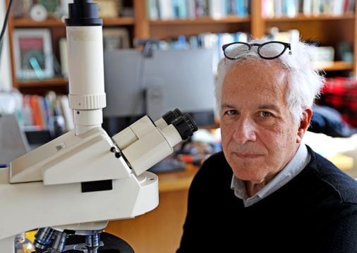
Adam Piore can be reached at adam.piore@globe.com.