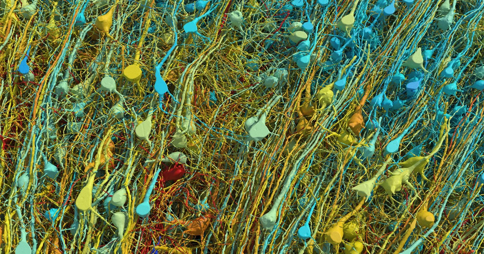
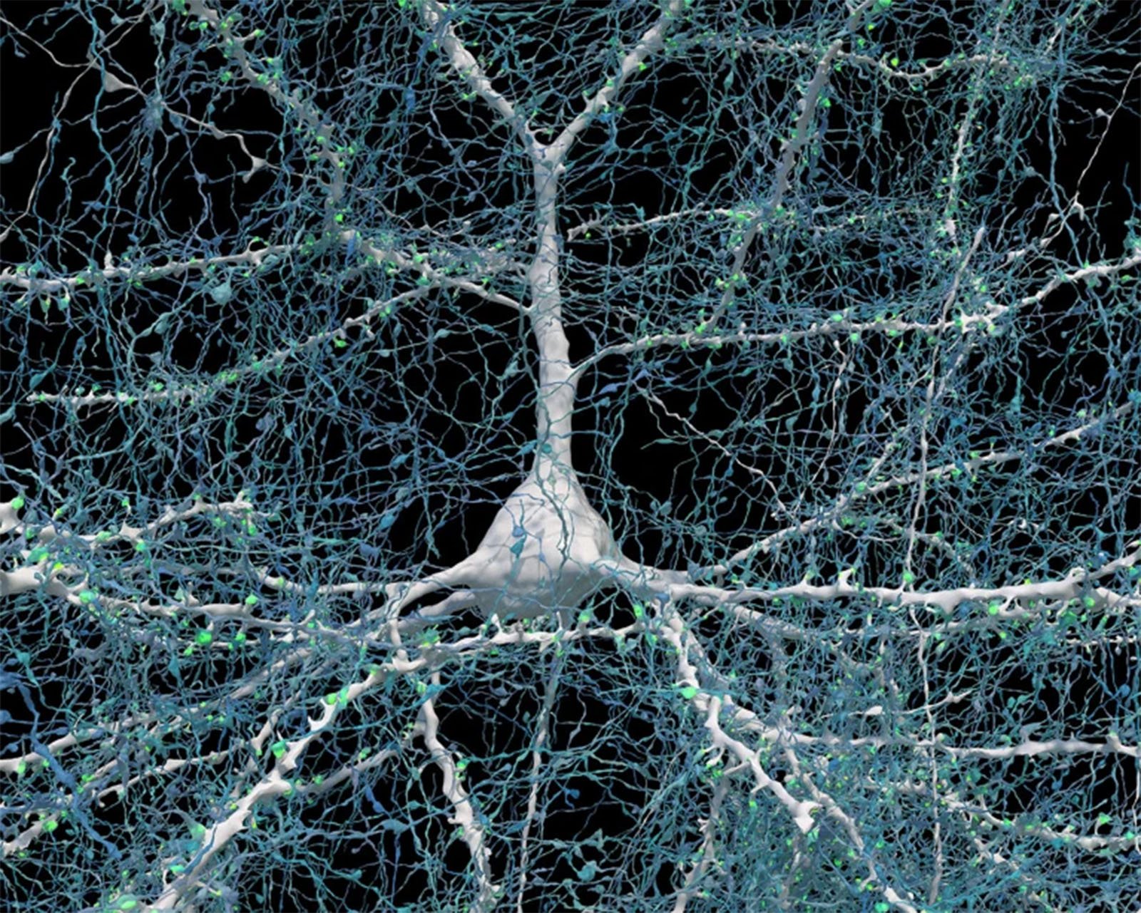
For something as central to all human existence and experience as the brain, many aspects of it remain completely mysterious. Thanks to revolutionary new digital images, models and 3D maps created by Google Research and scientists at Harvard University, the physical structure of the brain has never been so clear.
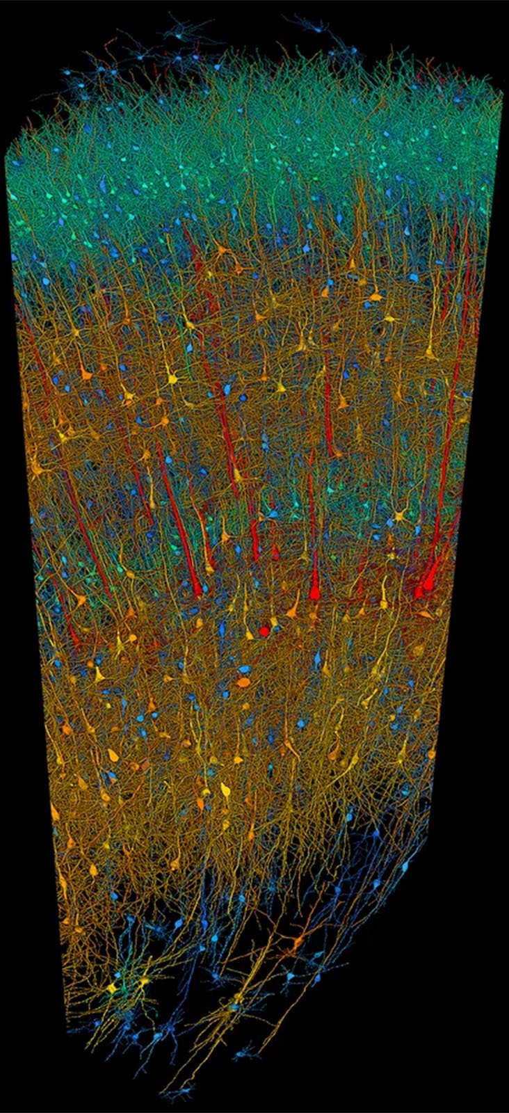
The technology in the game is remarkable. The team, consisting of researchers from Google and Harvard University’s Lichtman Lab, combined electron microscopy and cutting-edge artificial intelligence to inspect a tiny brain sample — just one cubic millimeter (0.000002 pint) — and create a litany of new images, models , and cards.
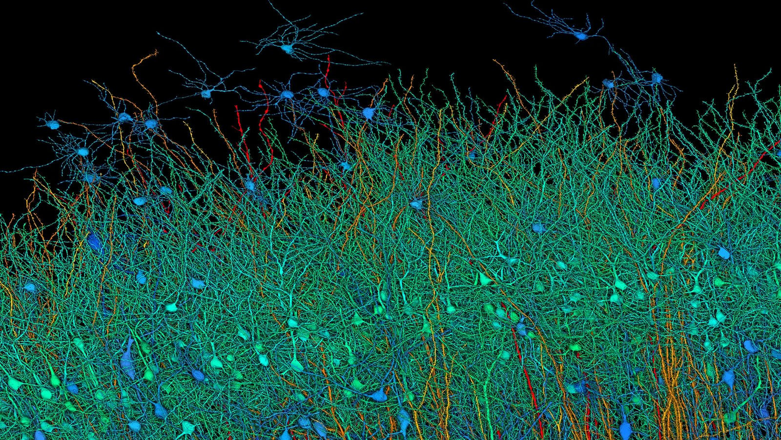
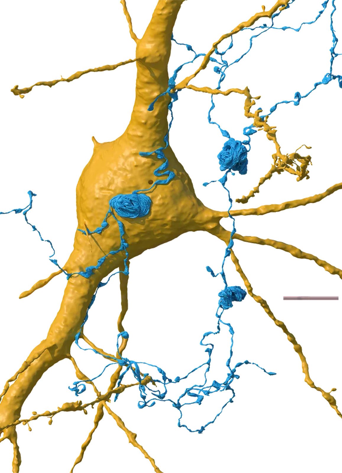
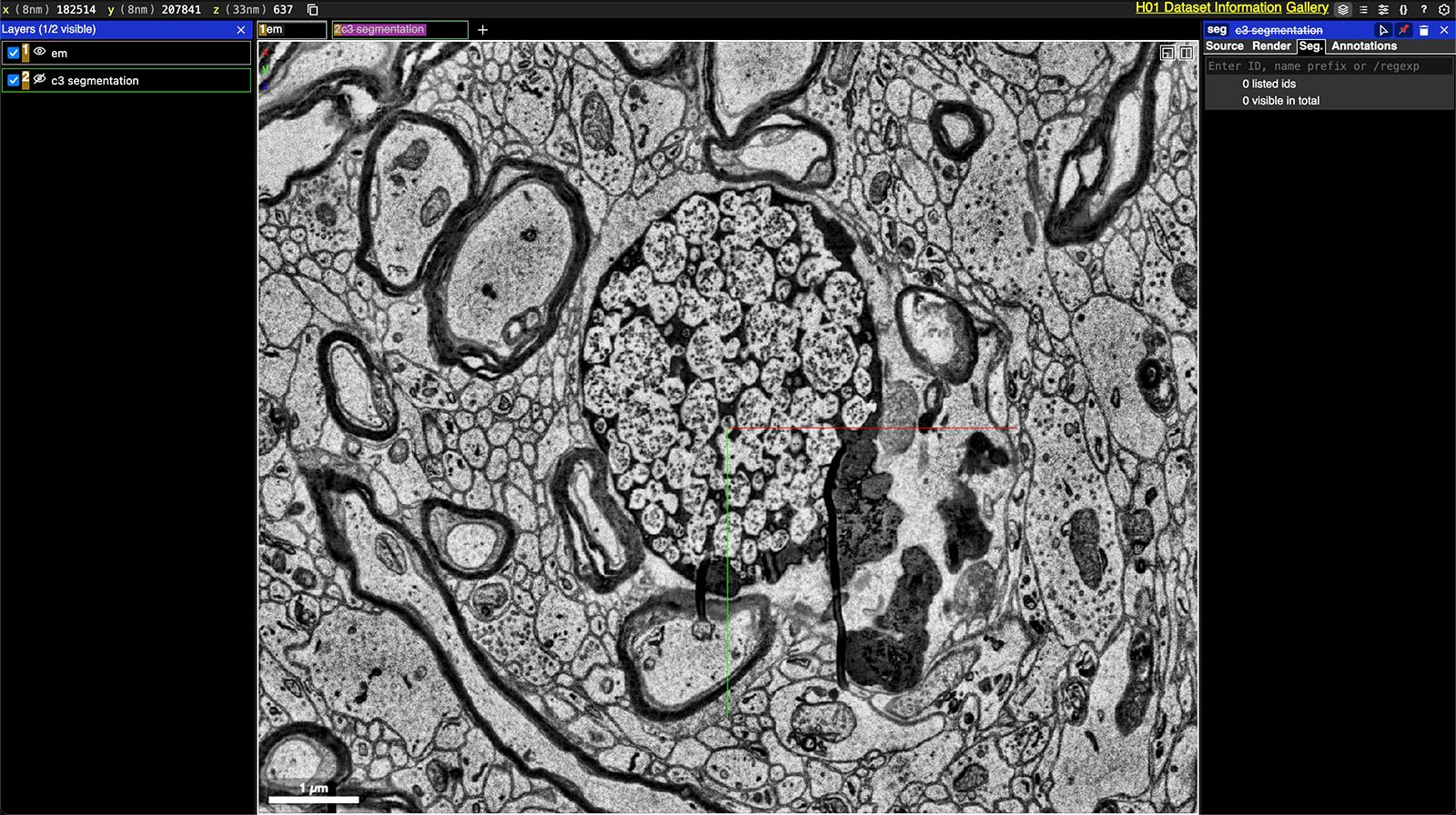
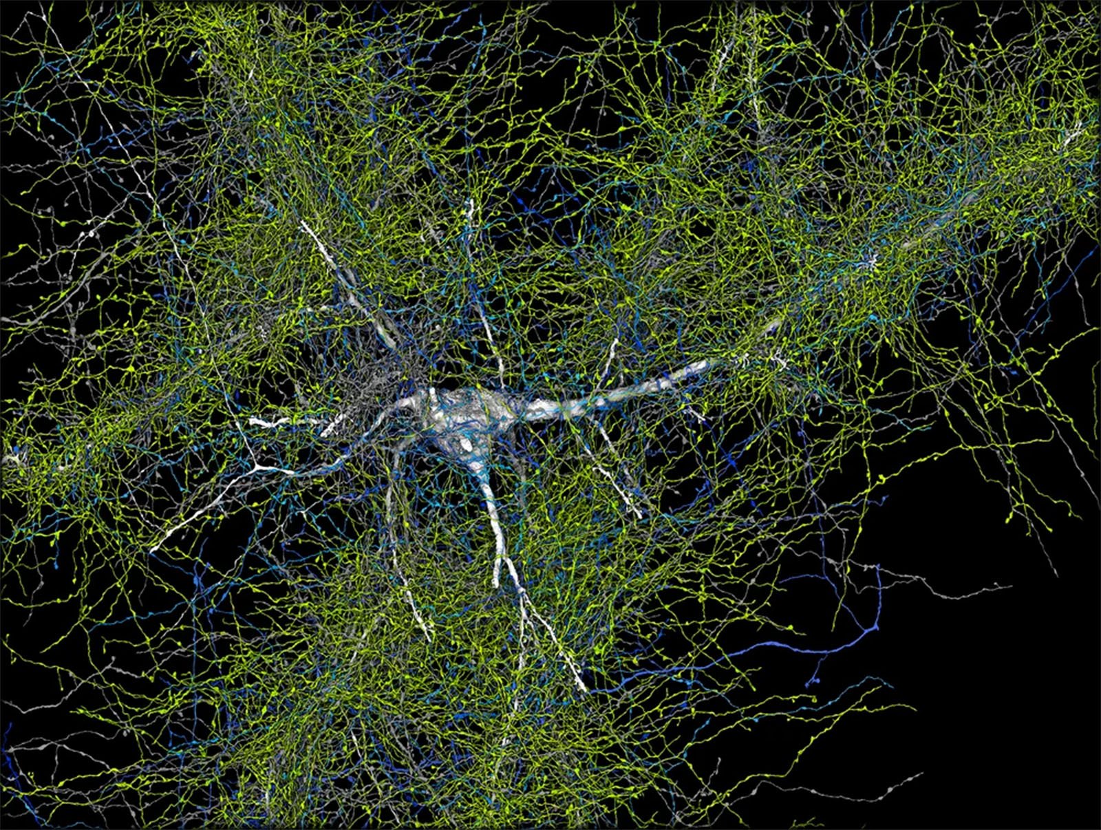
There are about 57,000 cells and 150 million synapses, the vital connections between neurons, in this single cubic millimeter of brain. The initial incision was made by a 45-year-old woman during a surgical procedure to treat epilepsy, and this small piece of brain was then cut into about 5,000 individual slices, each about 34 nanometers thick, so that they could be imaged individually using electron microscopy. And yes, this “brain delicacy” is quite remarkable in itself and useful for other brain research.
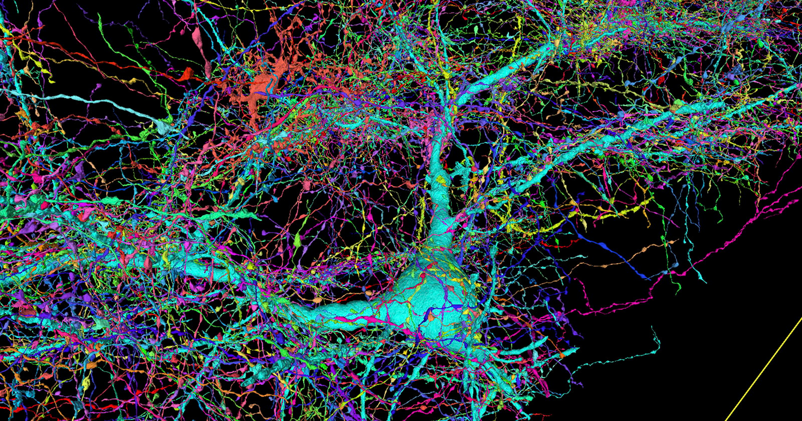
Neuroscientists Viren Jain of Google and Jeff Lichtman of Harvard—the namesake of the university’s Lichtman Lab—worked together, along with dozens of others, to take thousands of microscopic images of brain tissue and reconstruct them using custom AI models into an entire 3D sample. The project took about a decade to complete and includes 1.4 petabytes (1,400 terabytes) of data that is publicly available. Treating the entire human brain the same way would be one billion terabytes, which is almost as much digital data as the world creates in a year.
“It’s a little humbling,” Wein says of the project. “How are we ever going to come to terms with all this complexity?” The average adult brain is about 1,200 centimeters3or 1.2 million millimeters3. The decade-long project to reconstruct part of the brain looked at just 0.000083 percent of a typical adult’s brain.
“The word ‘fragment’ is ironic,” says Lichtman. “One terabyte to most people is gigantic, but a fragment of a human brain – just one tiny, tiny bit of a human brain – is still thousands of terabytes.”
This type of work constituted Lichtman’s entire illustrious career. He is a specialist in the burgeoning field of “connectomics,” which is like genomics, but for the brain. Lichtman and his team are working to create complete, detailed images of all brain structures at the single-cell level. The goal is, by making this map, to gain critical insight into brain function and related pathology.
The initial stage of this grand project is already progressing. The team found a small number of axons formed in a strange swirling pattern unlike anything seen before. However, given that the small part of the brain was taken from a woman with epilepsy, it is unclear whether this spiral was a result of her illness.
Last year, Lichtman called the brain “depressingly complex” in an interview with Journal of Harvard. However, given the progress he and his team have made, he has reason for some optimism.
“If we get to a point where working with a whole mouse brain becomes routine, you could consider doing it in, say, animal models of autism.” There is a level of understanding about the brain that does not currently exist. We know about the outward manifestations of behavior. We know of some of the molecules that are disordered. But between them, the wiring diagrams, until now there was no way for me to see them. Now there is a way,” Lichtman explained.
Image Credits: Google Research and Lichtman Lab, Harvard University. Translations by D. Berger. The full data release is available via Google, and the research is detailed in a paper published in Science.