Sign up for CNN’s Wonder Theory science newsletter. Explore the universe with news of fascinating discoveries, scientific breakthroughs and more.
CNN
—
Ten years ago, Dr. Jeff Lichtman, a professor of molecular and cellular biology at Harvard University, received a small brain sample in his laboratory.
Although small, 1 cubic millimeter of tissue was large enough to contain 57,000 cells, 230 millimeters of blood vessels and 150 million synapses.
“It was smaller than a grain of rice, but we started cutting it up and looking at it and it was really beautiful,” he said. “But as we accumulated the data, I realized that we just had way more than we could handle.”
In the end, Lichtman and his team came up with 1,400 terabytes of data from the sample—roughly the contents of more than 1 billion books. Now, after a decade of close collaboration by the lab team with Google scientists, this data has become the most detailed map of a human brain sample ever created.
The brain sample comes from a patient with severe epilepsy. It’s standard procedure, Lichtman said, to remove a small part of the brain to stop the seizures and then look at the tissue to make sure it’s normal. “But it was anonymized, so I knew next to nothing about the patient other than their age and gender,” he said.
To analyze the sample, Lichtman and his team first cut it into thin sections using a knife with a blade made of diamond. The sections were then embedded in hard resin and sliced again, very thinly. “About 30 nanometers, or roughly 1,000 of the thickness of a human hair. They were virtually invisible if it weren’t for the fact that we had stained them with heavy metals, which made them visible when taking electronic images,” he said.
The team ended up with several thousand slices, which were taken with a custom-made tape, creating something like a film strip: “If you take a picture of each of these sections and align those pictures, you get a three-dimensional slice of the brain at the microscopic level.
The researchers then realized they needed help with the data, as the resulting images would take up a significant amount of storage space.
Lichtman knew that Google was working on a digital map of the fruit fly brain, published in 2019, and had the right computer hardware for the job. He contacted Viren Jain, a senior scientist at Google who was working on the fruit fly project.
“There were 300 million individual images (in the Harvard data),” Jain said. “What makes it so great is that you’re imaging at very high resolution, the level of an individual synapse. And in this small sample of brain tissue alone, there were 150 million synapses.
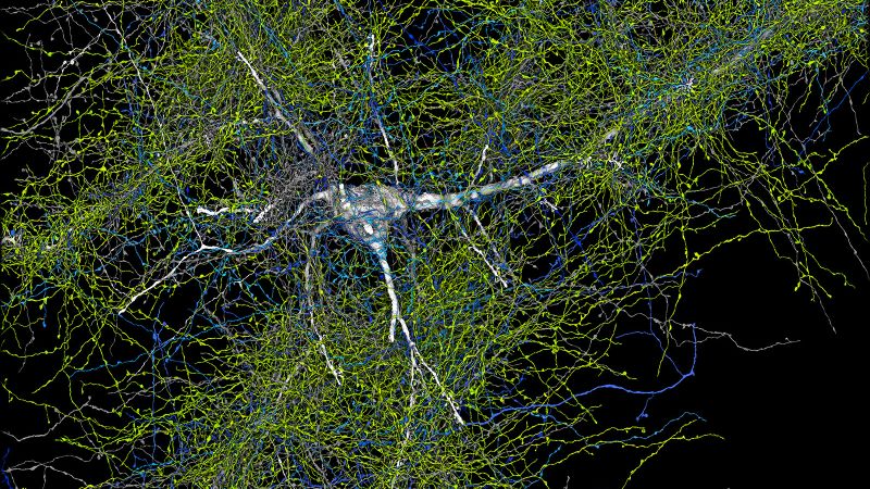
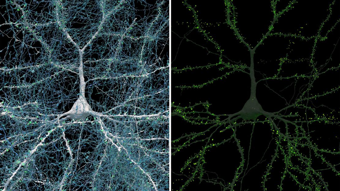
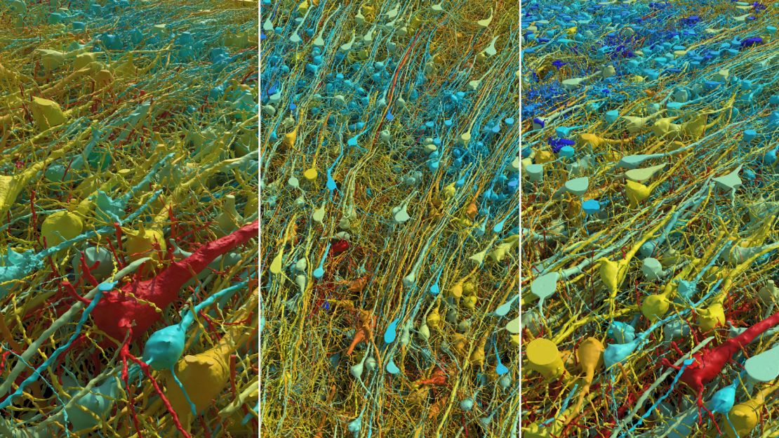
To make sense of the images, scientists from Google uses artificial intelligence-based processing and analysis, identifying what type of cells are in each photo and how they are connected. The result is an interactive 3D model of brain tissue and the largest dataset ever made at this resolution of a human brain structure. Google made it available online as “Neuroglancer,” and a study was published in the journal Science at the same time, with Lichtman and Jain among the co-authors.
Understanding the brain
A collaboration between the Harvard and Google teams resulted in colorful images that make individual components more visible, but are otherwise a faithful representation of the tissue.
“The colors are completely arbitrary,” Jane explained, “but other than that, there’s not a lot of artistic freedom here. The whole point of this is that we’re not making this up – these are the real neurons, the real wiring that exists in this brain, and we’re really making it convenient and accessible for biologists to look at and study.
The data contained some surprises. For example, instead of forming a single connection, pairs of neurons instead have more than 50. “It’s kind of like if two houses on a block have 50 separate phone lines connecting them. What’s going on? Why are they so strongly related? We don’t yet know what the function or significance of this phenomenon is, we will have to study it further,” he said.
Ultimately, observing the brain at this level of detail could help researchers make sense of unsolved medical conditions, according to Lichtman.
“What does it mean to understand our brain? The best we can do is describe it and hope that from these descriptions will come an awareness of, for example, how normal brains are different from brains that are disordered, in adult psychiatric illness or developmental disorders like the autism spectrum – that kind of comparison will be very valuable,” he said. “Ultimately, it will give us some insight into what’s wrong, which in most cases we’re still in the dark about.”
Lichtman also believes the dataset may be full of other amazing details that, because of its size, have yet to be discovered: “And that’s why we’re sharing it online, so anyone can look at it and find things,” he added. .
The team behind the project then aims to create a complete map of the mouse brain, which would require between 500 and 1,000 times more data than a human brain sample.
“That would mean 1 exabyte, which is 1,000 petabytes,” Lichtman said. “A lot of people are thinking hard about how we’re going to do this, and we’re in the first year of a five-year proof-of-principle. I think it would be a watershed moment for neuroscience, to have a complete wiring diagram of the mammalian brain; will answer many, many questions. And of course, it will reveal many more problems, things that we did not expect.
How about mapping an entire human brain? That would be another 1,000 times larger, Lichtman explained, meaning the data would amount to 1 zettabyte. In 2016, that was the amount of all Internet traffic for the year, according to Cisco. Right now, Lichtman said, not only would it be difficult to even store that much data, but there would be no ethically acceptable way to find a pristine, well-preserved human brain.
Researchers in the same field who were not involved in the work expressed their enthusiasm when contacted by CNN for comment.
“This study is wonderful, and there is so much to learn from data like this,” said Michael Bienkowski, assistant professor of physiology and neurology at the University of Southern California Keck School of Medicine.
“Much of what we think we understand about the human brain has been extrapolated from animals, but research like this is critical to uncovering what really makes us human. “Visualizing neurons and other brain cells is really challenging because of their sheer density and complexity, and the current dataset doesn’t capture the longer-range connections,” Bienkowski said.
“What other areas of the brain do these inputs come from, and where do the outputs go after they leave the area?” But to see all these different types of cells and their interactions is amazing and makes you appreciate what a masterpiece of architecture life has given us,” he added.
Andreas Tolias, a professor of ophthalmology at Stanford University in California, agreed. “This is a remarkable technical study that reconstructs the structure of the human cortex at high resolution,” he said. “I was particularly excited by the discovery of rare axons capable of forming up to 50 synapses. This finding is intriguing and raises important questions about their computational roles.
The brain mapping project opens the door to future research, according to neuroscientist Olaf Sporns.
“Each human brain is a vast network of billions of nerve cells,” said Sporns, Indiana University Distinguished Professor of Psychology and Brain Sciences. “This network allows cells to communicate in very specific patterns that are fundamental to memory, thought and behavior. Mapping this network, the human connectome, is critical to understanding how the brain works,” he added, noting that the study breaks new ground toward this important goal and offers exciting new opportunities for research and discovery.