Sign up for CNN’s Wonder Theory science newsletter. Explore the universe with news of fascinating discoveries, scientific breakthroughs and more.
CNN
—
An extinct ribbon-like sea creature about the size of a human hand was one of the earliest animals to develop a precursor to a backbone. Scientists recently identified the animal’s nerve cord by twisting it inside out. They turned his fossils upside down.
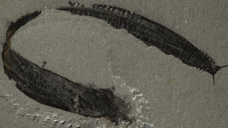
Paleontologist Charles Doolittle Walcott first encountered Pikaia fossils in the Burgess Shale deposits of British Columbia, dating back 508 million years ago, and described them in a 1911 treatise. The animal was approximately 6.3 inches (16 centimeters) long ) and had a flattened, curved body and a small head topped by two tentacles and surrounded by external gills. These were originally thought to be rudimentary legs, so the animal was positioned with these structures facing downwards.
In 2012, after decades of studying Pikaia fossils, researchers described its fossilized internal structures in great detail. They identified a long strand near the abdomen as a blood vessel and called a three-dimensional sausage-shaped structure running under the animal’s back a dorsal organ, possibly used for internal support, although such an organ was anatomically unlike anything seen in fossils or live animals.
However, a recent analysis of Pikaia fossils by another team of scientists, published June 11 in the journal Current Biology, overturned that view and all other earlier studies of Pikaia.
According to the researchers, earlier anatomical interpretations placed the animal wrong side up. The so-called dorsal organ was actually located in the abdomen and was the Pikaia’s gut. The supposed blood vessel is a nerve cord, a feature associated with the group of animals known as chordates, of the phylum Chordate.
Giovanni Mussini
Annotated images show the newly reworked organization of Pikaia gracilens. Abbreviations in field C indicate key features in the fossil seen in field B: Pikaia head tentacles (Tc); dorsal nerve cord (In); dorsal nerve cord (Nc); possible gonads (?Go); and the myosepta, or connective fascia (Ms). The drawing in field G identifies the features of the fossa in field F: anterior processes (Aa); pharyngeal cavity (Ph); intestinal channel (Gu); and myomeres or muscle segments (My). Fossil specimens are from the Smithsonian National Museum of Natural History, except for the fossils in Box I from the Royal Ontario Museum.
All chordates, such as vertebrates, eel-like lancelets, and shellfish or sea squirts, at some point in their lives have a flexible, rod-like nerve structure called the notochord in their back.
Pikaia was originally thought to be a worm, then later upgraded to an early type of chordate, based on features such as the shape of certain muscles and the position of the anus. But experts weren’t sure exactly where Pikaia belonged in the chordate family tree.
With the description of a nerve cord, Pikaia can now be considered part of the founding lineage of all chordates, although there are no direct descendants alive today, the study authors report.
Pikaia’s reversal “clears things up a lot,” said evolutionary biologist Dr. John Mallat, a clinical professor at the University of Idaho. Mallatt, who was not involved in the new research, published a paper on Pikaia in 2013, working from the established (and upside down) body position.
In retrospect, the truth is “hiding in plain sight,” and the reversal in orientation resolves questions about why the putative blood vessel and dorsal structure of Pikaia clashed with the established anatomical features of other chordates, Mallatt said.
“Pikaia suddenly became a lot less weird,” he said.
The reassessment of which way is up for Pikaia arose years ago with co-author of the new study, Dr. Jakob Winter, a professor of macroevolution at the University of Bristol in the United Kingdom, said lead study author Giovanni Mussini, a researcher and doctoral student in the Department of Earth Sciences. at the University of Cambridge in the UK.
There were a number of reasons for revising earlier interpretations of the fossils, Mussini told CNN. First, there was the enigma of what scientists believed was the position of the dorsal organ. Its placement – near what is thought to be Pikaya’s back – seems to rule out the possibility that the organ is an intestine.
Once Pikaia was turned upside down, however, the location and characteristics of the organ made more sense from an anatomical point of view. It expands and extends into the animal’s pharynx, the area of the throat where the intestines normally connect to the mouth. Its 3D status can be explained by the presence of chemically reactive tissues – hallmarks of the intestine. In other Burgess Shale fossils, the abundance of ions and reactive compounds normally found in intestinal tissue cause the digestive structures to mineralize faster than the rest of the body and thus retain more of their original forms. The structures in Pikaia’s organ are likely the remains of ingested food, according to the study.
Giovanni Mussini
An image of a fossil specimen of Pikaia in the Smithsonian’s National Museum of Natural History shows the intestinal canal, blocks of muscle tissue known as myomeres, and the dorsal nerve cord. A light-colored sediment is visible inside the intestine (toward the head on the right).
In an inverted Pikaia, the outer gills that previously pointed downwards were now pointed upwards, as are the outer gills of modern mudskippers and axolotls.
Pikaia’s reversal also changed the orientation of the muscle groups that come together in a wave-like formation. These muscles, called myomeres, are a key feature in vertebrates. In Pikaia’s new position, the strongest point of flexion of these muscles is along its back, which is also true of the arrangement of myomeres in other vertebrates.
“This makes the movement of Pikaia compatible with what we see in modern chordates,” Mussini said.
The putative blood vessel of Pikaia was also anatomically puzzling, as it lacked the branches normally found in vertebrate blood vessels.
“It’s one line going through most of the body to the head where it splits into these two strands in the tentacles,” Mussini said.
Giovanni Mussini
An interpretive drawing of the head of Pikaia gracilens from a fossil specimen in the Smithsonian National Museum of Natural History highlights a thickened portion of the dorsal nerve cord. The discovery of other Cambrian fossilized nervous systems has helped scientists take a new look at how Pikaia was organized.
An important part of identifying the structure as a nerve cord was fossilized nervous systems in other animals from the Cambrian period (541 million to 485.4 million years ago) that have been discovered in the past decade, Mussini added.
“We understand better how nerve cords and other tissues fossilize because we’ve been fortunate enough to find quite a few Cambrian nervous systems preserved in other deposits,” he said, “mostly from Chinese fossils that have come to light in the last few years.”
Many of these fossils are arthropods—invertebrates with exoskeletons—with living relatives such as insects, arachnids, and crustaceans; comparing fossils to modern arthropods helped paleontologists identify preserved internal tissues. One example is a fossil specimen of the Cambrian arthropod Mollisonia, which shows a brain organization comparable to that of living spiders, scorpions and horseshoes, Mussini said.
Although there are no living analogues of Pikaia, fossil arthropod data have given scientists a more detailed frame of reference for Pikaia’s nerve cord. Like other fossilized nerve tissues, the nerve cord in Pikaia was dark, carbon-rich, and relatively fragile compared to other fossilized tissues.
This nerve cord cements Pikaia’s status as a chordate, placing it “pretty much at the core of what we would consider traditional chordates,” Mallat said.
Much of Pikaia’s anatomy remains a mystery, but looking at it from a new angle could offer fresh insights into its puzzling array of functions, Mussini said.
“Many of these details have only come to light in the last 10 or 12 years,” Mussini added. “The authors of the 2012 paper could certainly be forgiven for not bringing these details into the conversation because it is a work in progress.”
Mindy Weisberger is a science writer and media producer whose work has been published in Live Science, Scientific American, and How It Works magazine.